Dominant Frequency (DF) is a term used to identify the frequency related with the greatest power/amplitude in the signal.
In order to understand DF, it is good to start with Fast Fourier Transform spectrum (FFT). FFT spectrum is basically a plot showing contributions of various frequencies in input signal. For example, if we would start with a signal composed of a low frequency, slow oscillation and, on top of it, low amplitude fast oscillations, we would get a following FFT spectrum:
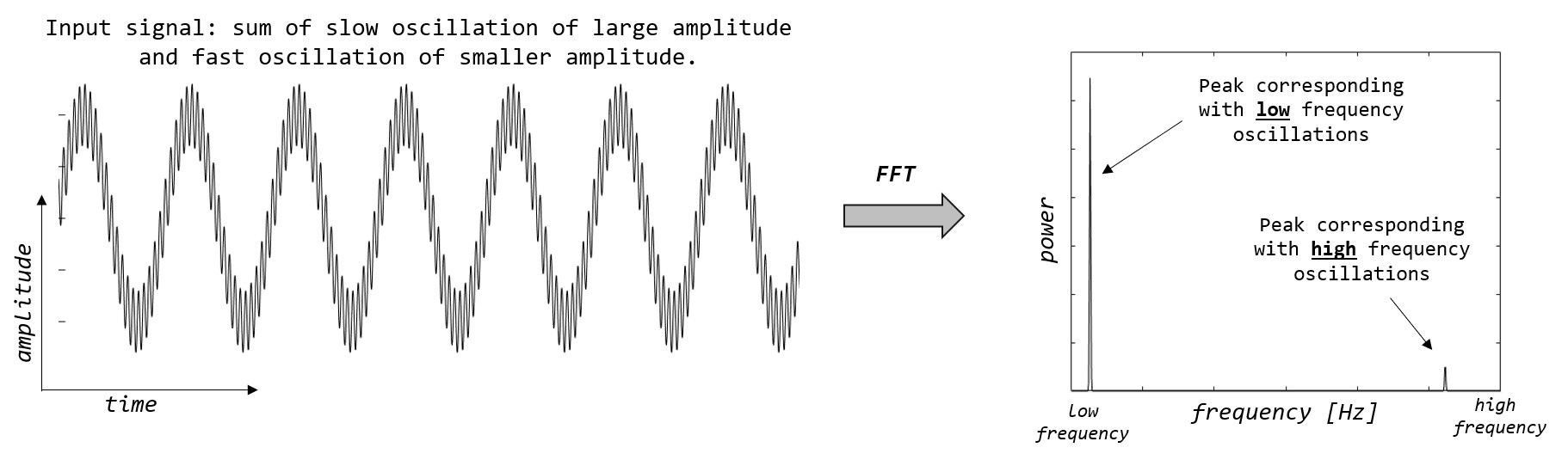
In resultant FFT spectrum, we find two narrow peaks, corresponding with both oscillations in our input signal (slow, weaving oscillation and superimposed fast one). So, simply, FFT shows us which frequencies are present in original signal.
It is also worth to notice, that height of the peak corresponds with the amplitude of the oscillation of given frequency. Our low frequency oscillation has much greater amplitude in input signal, and that’s why it has also much greater amplitude (power) in FFT spectrum.
In cardiac electrophysiology, FFT analysis are often used in context of analysis of Atrial Fibrillation (AF) electrograms. During AF, we see a variability in electrograms, but we also notice rather stable underlying cycle length (at least in range of few seconds).
What would we see if we applied FFT to AF electrograms?
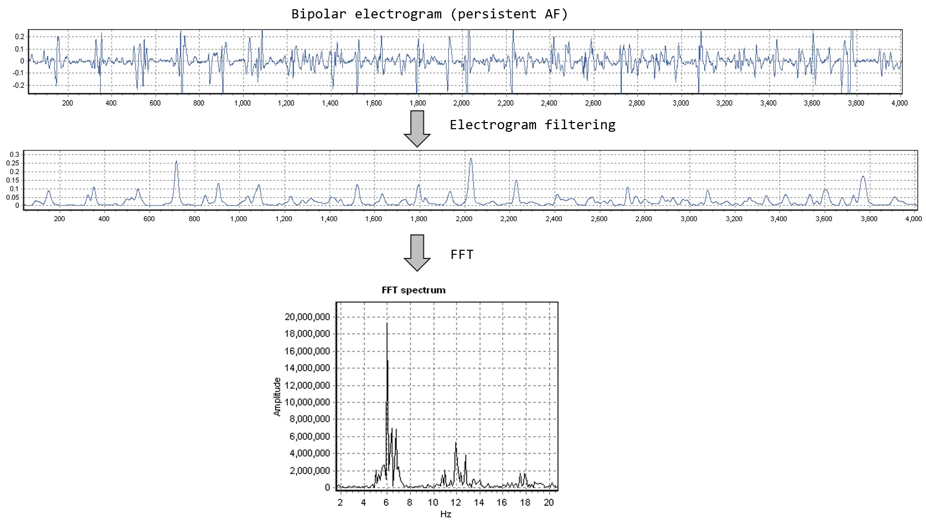
Note: before applying FFT to bipolar electrogram, filtering steps are usually performed to remove very high frequency noise and baseline wandering. Typical filtering steps: Butterworth band-pass 40-250 Hz -> Rectification -> Butterworth low pass 20 Hz (1).
As we see, FFT spectrum contains two peaks. One around 6 Hz (which corresponds with 167 ms cycle length) and one around 12 Hz. The first peak we call dominant frequency, because it has the greatest amplitude and is within the physiological range. The second peak is disregarded because of two reasons: (i) it is a multiplication of the base peak (12 Hz = 2×6 Hz) – which is called a harmonic and is actually an ‘side effect’ artifact of FFT analysis), (ii) it is in non-physiological range (it would correspond with cycle length of 83 ms which is below typical short refractory period).
Of interest, DF was used to guide catheter ablation of AF with promising results (2,3). It has not been widely adopted but is undergoing constant refinement (4).
The strength of the FFT analysis, is that we can obtain an estimate of underlying cycle length without annotating local activations. The weakness is that DF is just an estimate of the cycle length – sort of average value – which actually may never be found if we would look at intervals between consecutive activations.
FFT analysis of AF electrograms should be performed with great caution and consideration. Elvan et. al showed in their analysis that DF correlates poorly with manually annotated mean AF cycle length (5). However, even such detailed analysis attracted critique pointing to technical complexity of FFT analysis and influence of electrogram fractionation (6,7) that can confound both FFT analysis and activation annotation.
References:
1. Everett TH, Lai-Chow Kok, Vaughn RH, Moorman R, Haines DE. Frequency domain algorithm for quantifying atrial fibrillation organization to increase defibrillation efficacy. IEEE Transactions on Biomedical Engineering. 2001 Sep;48(9):969–78.
2. Sanders Prashanthan, Berenfeld Omer, Hocini Mélèze, Jaïs Pierre, Vaidyanathan Ravi, Hsu Li-Fern, et al. Spectral Analysis Identifies Sites of High-Frequency Activity Maintaining Atrial Fibrillation in Humans. Circulation. 2005 Aug 9;112(6):789–97.
3. Pachon M JC, Pachon M EI, Pachon M JC, Lobo TJ, Pachon MZ, Vargas RNA, et al. A new treatment for atrial fibrillation based on spectral analysis to guide the catheter RF-ablation. Europace. 2004 Nov;6(6):590–601.
4. Gadenz L, Hashemi J, Shariat MH, Gula L, Redfearn DP. Clinical Role of Dominant Frequency Measurements in Atrial Fibrillation Ablation – A Systematic Review. J Atr Fibrillation [Internet]. 2017 Apr 30 [cited 2019 Sep 29];9(6). Available from: https://www.ncbi.nlm.nih.gov/pmc/articles/PMC5673341/
5. Elvan A, Linnenbank AC, van Bemmel MW, Misier ARR, Delnoy PPHM, Beukema WP, et al. Dominant frequency of atrial fibrillation correlates poorly with atrial fibrillation cycle length. Circ Arrhythm Electrophysiol. 2009 Dec;2(6):634–44.
6. Berenfeld O, Jalife J. Letter by Berenfeld and Jalife Regarding Article “Dominant Frequency of Atrial Fibrillation Correlates Poorly With Atrial Fibrillation Cycle Length.” Circ Arrhythm Electrophysiol. 2010 Feb;3(1):e1–3.
7. Platonov PG, Stridh M, Sörnmo L. Letter by Platonov et Al regarding article, “Dominant frequency of atrial fibrillation correlates poorly with atrial fibrillation cycle length.” Circ Arrhythm Electrophysiol. 2010 Apr;3(2):e4; author reply e5.